Temporal Lobe Hsv Encephalitis Mri - Medpix Case Herpes Encephalitis Hsv 1 / But changes are not specific for hsv (e.g.
Get link
Facebook
X
Pinterest
Email
Other Apps
Temporal Lobe Hsv Encephalitis Mri - Medpix Case Herpes Encephalitis Hsv 1 / But changes are not specific for hsv (e.g.. Mri findings in acute encephalitis: In addition, it was observed in various pathological conditions: Key imaging features include bilateral or unilateral signal abnormality in the temporal lobes that extends to the limbic system, early hemorrhagic changes, restriction on dwi, and abnormal enhancement. It is usually bilateral but asymmetrical. In the present study, flair bth was most frequently seen in patients with hsv encephalitis.
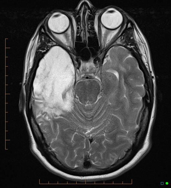
A 40‑year‑old male patient with herpes simplex virus encephalitis presented with altered sensorium. The differential diagnoses include limbic encephalitis (paraneoplastic), gliomatosis cerebri, and status epilepticus. Herpes simplex (hsv) encephalitis is the most common cause of fatal sporadic fulminant necrotizing viral encephalitis and has characteristic imaging findings. Severe edema, petechial hemorrhage, and massive hemorrhagic tissue necrosis can be seen on gross specimens. Two subtypes are recognized which differ in demographics, virus, and pattern of involvement.
Herpes Simplex Type 1 Encephalitis Eurorad from www.eurorad.org Severe edema, petechial hemorrhage, and massive hemorrhagic tissue necrosis can be seen on gross specimens. Two subtypes are recognised which differ in demographics, virus, and pattern of involvement. In the present study, flair bth was most frequently seen in patients with hsv encephalitis. 11 public playlist includes this case Although far less common, essentially any part of the central nervous system can be involved 9. Mri revealed hyperintensities on t2 weighted images in both frontal and temporal lobes suggestive of herpes simplex encephalitis (fig 1). Metabolic, vascular, neoplastic, infective and paraneoplastic diseases. Magnetic resonance imaging (mri) revealed a newly identified enhancing left sphenoid wing meningioma with peritumoral edema in the underlying brain (figure 1).
We describe the spectrum of etiologies associated with temporal lobe (tl) encephalitis and identify clinical and radiologic features that distinguish herpes simplex encephalitis (hse) from its mimics.
Watershed distribution ischemia in areas remote from the primary herpetic lesions may be seen. Herpes simplex encephalitis typically manifests in older adults (about 50% of cases) with headache, fever, altered sensorium, and even seizure. Hippocampus, temporal lobe, and the ponsfigure 4a.7. Herpes simplex virus (hsv) is the most common cause of acute fatal sporadic encephalitis, with a particular predilection for the limbic system. Unfortunately, the imaging findings are often nonspecific with overlapping appearances. This lesion was seen in all patients as the disease progressed. Severe edema, petechial hemorrhage, and massive hemorrhagic tissue necrosis can be seen on gross specimens. He was managed with intravenous acyclovir 30mg/kg and supportive therapy. that appeared in a recent issue of clinical infectious diseases. However, mimics of hse, including other infections and increasingly recognized autoimmune causes, have been described in cases of tl encephalitis. But changes are not specific for hsv (e.g. The characteristic feature of hse is hemorrhagic necrosis of the temporal lobe. The basal ganglia are usually spared.
Herpes simplex (hsv) encephalitis is the most common cause of fatal sporadic fulminant necrotizing viral encephalitis and has characteristic imaging findings. Metabolic, vascular, neoplastic, infective and paraneoplastic diseases. A 40‑year‑old male patient with herpes simplex virus encephalitis presented with altered sensorium. Key imaging features include bilateral or unilateral signal abnormality in the temporal lobes that extends to the limbic system, early hemorrhagic changes, restriction on dwi, and abnormal enhancement. Mri revealed hyperintensities on t2 weighted images in both frontal and temporal lobes suggestive of herpes simplex encephalitis (fig 1).
Temporal Lobe Atrophy Post Herpes Simplex Encephalitis Radiology Case Radiopaedia Org from prod-images-static.radiopaedia.org The characteristic feature of hse is hemorrhagic necrosis of the temporal lobe. Severe edema, petechial hemorrhage, and massive hemorrhagic tissue necrosis can be seen on gross specimens. Because early diagnosis is possible in more cases of herpes simplex encephalitis (hse) as a result of the high sensitivity of mri, now widely available, a larger number of patients are receiving appropriate treatment with iv acyclovir. Herpes simplex encephalitis typically manifests in older adults (about 50% of cases) with headache, fever, altered sensorium, and even seizure. Herpes simplex virus (hsv) is the most common cause of acute fatal sporadic encephalitis, with a particular predilection for the limbic system. that appeared in a recent issue of clinical infectious diseases. It is usually bilateral but asymmetrical. This lesion was seen in all patients as the disease progressed.
However, mimics of hse, including other infections and increasingly recognized autoimmune causes, have been described in cases of tl encephalitis.
A feasibility study of quantifying longitudinal brain changes in herpes simplex virus (hsv) encephalitis using magnetic resonance imaging (mri) and stereology. In the present study, flair bth was most frequently seen in patients with hsv encephalitis. Mri is the diagnostic modality of choice abnormal in 90%; Herpes simplex virus (hsv) encephalitis hsv encephalitis (hsve) is the most common cause of infectious encephalitis (1); The basal ganglia are usually spared. Encephalitis, magnetic resonance imaging, meningoencephalitis, viral. that appeared in a recent issue of clinical infectious diseases. Although far less common, essentially any part of the central nervous system can be involved 9. 1 mri t2 weighted image, showing bilateral temporal lobe hyperintensities. However, mimics of hse, including other infections and increasingly recognized autoimmune causes, have been described in cases of tl encephalitis. Two subtypes are recognized which differ in demographics, virus, and pattern of involvement. Because early diagnosis is possible in more cases of herpes simplex encephalitis (hse) as a result of the high sensitivity of mri, now widely available, a larger number of patients are receiving appropriate treatment with iv acyclovir. Limbic encephalitis, mca ischaemia, tumours, effects of seizures) hyperintense t2 signal in the medial temporal lobes, inferior frontal lobes and insula basal ganglia are usually spared
Herpes simplex (hsv) encephalitis is the most common cause of fatal sporadic fulminant necrotizing viral encephalitis and has characteristic imaging findings. The characteristic feature of hse is hemorrhagic necrosis of the temporal lobe. Unfortunately, the imaging findings are often nonspecific with overlapping appearances. Although far less common, essentially any part of the central nervous system can be involved 9. He was managed with intravenous acyclovir 30mg/kg and supportive therapy.
Plos One A Feasibility Study Of Quantifying Longitudinal Brain Changes In Herpes Simplex Virus Hsv Encephalitis Using Magnetic Resonance Imaging Mri And Stereology from journals.plos.org We describe the spectrum of etiologies associated with temporal lobe (tl) encephalitis and identify clinical and radiologic features that distinguish herpes simplex encephalitis (hse) from its mimics. In addition, it was observed in various pathological conditions: A feasibility study of quantifying longitudinal brain changes in herpes simplex virus (hsv) encephalitis using magnetic resonance imaging (mri) and stereology. Encephalitis, magnetic resonance imaging, meningoencephalitis, viral. In the present study, flair bth was most frequently seen in patients with hsv encephalitis. that appeared in a recent issue of clinical infectious diseases. 1 mri t2 weighted image, showing bilateral temporal lobe hyperintensities. A brain biopsy was performed and the histology was consistent with encephalitis.
Mri findings in acute encephalitis:
It is usually bilateral but asymmetrical. This lesion was seen in all patients as the disease progressed. Two subtypes are recognised which differ in demographics, virus, and pattern of involvement. The lateral temporal lobe and insula are less commonly involved, whereas the basal ganglia, in contrast, are frequently involved, helpful in distinguishing it from hsv encephalitis which characteristically spares the basal ganglia 8. However, mimics of hse, including other infections and increasingly recognized autoimmune causes, have been described in cases of tl encephalitis. Severe edema, petechial hemorrhage, and massive hemorrhagic tissue necrosis can be seen on gross specimens. Because early diagnosis is possible in more cases of herpes simplex encephalitis (hse) as a result of the high sensitivity of mri, now widely available, a larger number of patients are receiving appropriate treatment with iv acyclovir. A feasibility study of quantifying longitudinal brain changes in herpes simplex virus (hsv) encephalitis using magnetic resonance imaging (mri) and stereology. In addition, it was observed in various pathological conditions: Limbic encephalitis, mca ischaemia, tumours, effects of seizures) hyperintense t2 signal in the medial temporal lobes, inferior frontal lobes and insula basal ganglia are usually spared He was managed with intravenous acyclovir 30mg/kg and supportive therapy. We describe the spectrum of etiologies associated with temporal lobe (tl) encephalitis and identify clinical and radiologic features that distinguish herpes simplex encephalitis (hse) from its mimics. Herpes simplex virus (hsv) is the most common cause of acute fatal sporadic encephalitis, with a particular predilection for the limbic system.
that appeared in a recent issue of clinical infectious diseases hsv encephalitis mri. (a) on presentation there may be unilateral or bilateral asymmetric involvement of limbic system structures, including the temporal lobes, insulae, and cingulate gyri.

Comments
Post a Comment