Hsv Encephalitis Mri Radiopaedia - Herpes Encephalitis Ct Scan Ct Scan Machine - It is a common finding on brain mri and a wide range of differentials should be considered 1.
Get link
Facebook
X
Pinterest
Email
Other Apps
Hsv Encephalitis Mri Radiopaedia - Herpes Encephalitis Ct Scan Ct Scan Machine - It is a common finding on brain mri and a wide range of differentials should be considered 1.. Two subtypes are recognized which differ in demographics, virus, and pattern of involvement. 10 public playlist includes this case The changes spare the basal ganglia, a feature which is helpful in distinguishing an mca infarct with hemorrhagic transformation from herpes simplex encephalitis. Radiopaedia is free thanks to our supporters and advertisers. The changes spare the basal ganglia, a feature which is helpful in distinguishing an mca infarct with hemorrhagic transformation from herpes simplex encephalitis, the diagnosis in this case.
For a general discussion, and for links to other system specific manifestations, please refer to the article on hydatid disease. Mri is the diagnostic modality of choice abnormal in 90%; It is reasonable to obtain an mri when patients are asymptomatic to ensure that no other abnormality is present which may be causing a recurrent chemical meningitis (e.g. Herpes simplex (hsv) encephalitis is the most common cause of fatal sporadic fulminant necrotising viral encephalitis and has characteristic imaging findings. Spinal hydatid disease is an uncommon manifestation of hydatid disease, caused by the larval stage of echinococcus granulosus, or less commonly e.
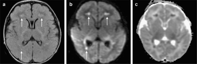
Diffusion Imaging In Brain Infections Radiology Key from radiologykey.com It is usually bilateral but asymmetrical. Hsv encephalitis | radiology case | radiopaedia.org. Note the high signal in the caudate heads and putamen on flair. Hypertrophic pachymeningitis is a condition where there is localized inflammatory thickening of the dura. For a general discussion, and for links to other system specific manifestations, please refer to the article on coccidioidomycosis. Radiopaedia is free thanks to our supporters and advertisers. Reference osborn a, et al. The changes spare the basal ganglia, a feature which is helpful in distinguishing an mca infarct with hemorrhagic transformation from herpes simplex encephalitis, the diagnosis in this case.
It is estimated to occur in ~2% of pati.
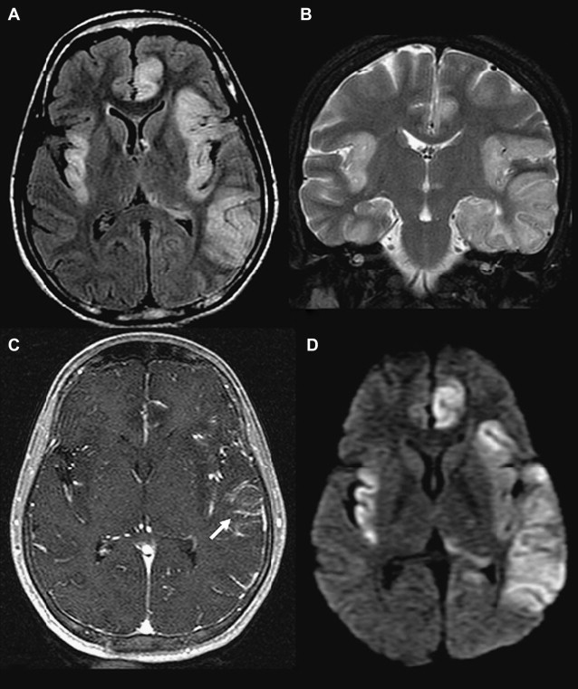
Two subtypes are recognized which differ in demographics, virus, and pattern of involvement. Become a gold supporter and see no ads. Bilateral temporal lobe t2 hyperintensity refers to hyperintense signal involving the temporal lobes on t2 weighted and flair imaging. Hypertrophic pachymeningitis is a condition where there is localized inflammatory thickening of the dura. Limbic encephalitis, mca ischaemia, tumours, effects of seizures) hyperintense t2 signal in the medial temporal lobes, inferior frontal lobes and insula basal ganglia are usually spared Reference osborn a, et al. Given the history of fever and seizures coupled with the mri findings of bilateral mesial temporal lobe changes, herpes encephalitis requires clinical consideration. The changes spare the basal ganglia, a feature which is helpful in distinguishing an mca infarct with hemorrhagic transformation from herpes simplex encephalitis, the diagnosis in this case. Mri demonstrates extensive edema in the right temporal lobe with areas of intrinsic high t1 signal, in keeping with hemorrhage. It is estimated to occur in ~2% of pati. The differential diagnoses include limbic encephalitis (paraneoplastic), gliomatosis cerebri, and status epilepticus. It is reasonable to obtain an mri when patients are asymptomatic to ensure that no other abnormality is present which may be causing a recurrent chemical meningitis (e.g. Axial t2 prominent swelling, increase t2 signal involving the left temporal lobe and insular cortex.
Axial t2 prominent swelling, increase t2 signal involving the left temporal lobe and insular cortex. Two subtypes are recognised which differ in demographics, virus, and pattern of involvement. Multilocularis.the larval stage is the cause of hydatid disease in humans 1. Cerebral malaria is a rare intracranial complication of a malarial infection. The changes spare the basal ganglia, a feature which is helpful in distinguishing an mca infarct with hemorrhagic transformation from herpes simplex encephalitis, the diagnosis in this case.
Neuroimaging Of Herpesvirus Infections In Children Springerlink from media.springernature.com It is reasonable to obtain an mri when patients are asymptomatic to ensure that no other abnormality is present which may be causing a recurrent chemical meningitis (e.g. Multilocularis.the larval stage is the cause of hydatid disease in humans 1. The changes spare the basal ganglia, a feature which is helpful in distinguishing an mca infarct with hemorrhagic transformation from herpes simplex encephalitis, the diagnosis in this case. The differential diagnoses include limbic encephalitis (paraneoplastic), gliomatosis cerebri, and status epilepticus. It is estimated to occur in ~2% of pati. 10 public playlist includes this case Spinal hydatid disease is an uncommon manifestation of hydatid disease, caused by the larval stage of echinococcus granulosus, or less commonly e. This patient went on to have hsv encephalitis proven on csf pcr.
Herpes simplex encephalitis typically manifests in older adults (about 50% of cases) with headache, fever, altered sensorium, and even seizure.
Mri demonstrates extensive edema in the right temporal lobe with areas of intrinsic high t1 signal, in keeping with hemorrhage. Become a gold supporter and see no ads. For a general discussion, and for links to other system specific manifestations, please refer to the article on coccidioidomycosis. This patient went on to have hsv encephalitis proven on csf pcr. This patient went on to have hsv encephalitis proven on csf pcr. Spinal hydatid disease is an uncommon manifestation of hydatid disease, caused by the larval stage of echinococcus granulosus, or less commonly e. Given the history of fever and seizures coupled with the mri findings of bilateral mesial temporal lobe changes, herpes encephalitis requires clinical consideration. Multilocularis, and describes a spectrum of disease involving the spinal cord, the spine, or both. Herpes simplex encephalitis radiology case radiopaedia mri demonstrates extensive edema in the right temporal lobe with areas of intrinsic high t1 signal, in keeping with hemorrhage. Hsv encephalitis | radiology case | radiopaedia.org. Two subtypes are recognised which differ in demographics, virus, and pattern of involvement. It is estimated to occur in ~2% of pati. Reference osborn a, et al.
Two subtypes are recognised which differ in demographics, virus, and pattern of involvement. Mri is the diagnostic modality of choice abnormal in 90%; A brain biopsy was performed and the histology was consistent with encephalitis. Reference osborn a, et al. Radiopaedia is free thanks to our supporters and advertisers.
Clinico Radiological Spectrum Of Bilateral Temporal Lobe Hyperintensity A Retrospective Review Abstract Europe Pmc from europepmc.org Herpes simplex (hsv) encephalitis is the most common cause of fatal sporadic fulminant necrotising viral encephalitis and has characteristic imaging findings. The differential diagnoses include limbic encephalitis (paraneoplastic), gliomatosis cerebri, and status epilepticus. The basal ganglia are usually spared. Note the high signal in the caudate heads and putamen on flair. Bilateral temporal lobe t2 hyperintensity refers to hyperintense signal involving the temporal lobes on t2 weighted and flair imaging. It is reasonable to obtain an mri when patients are asymptomatic to ensure that no other abnormality is present which may be causing a recurrent chemical meningitis (e.g. For a general discussion, and for links to other system specific manifestations, please refer to the article on hydatid disease. Radiopaedia is free thanks to our supporters and advertisers.
10 public playlist includes this case
Axial t2 prominent swelling, increase t2 signal involving the left temporal lobe and insular cortex. It is estimated to occur in ~2% of pati. Become a gold supporter and see no ads. This patient went on to have hsv encephalitis proven on csf pcr. A brain biopsy was performed and the histology was consistent with encephalitis. The differential diagnoses include limbic encephalitis (paraneoplastic), gliomatosis cerebri, and status epilepticus. Multilocularis.the larval stage is the cause of hydatid disease in humans 1. It is usually bilateral but asymmetrical. Spinal hydatid disease is an uncommon manifestation of hydatid disease, caused by the larval stage of echinococcus granulosus, or less commonly e. Limbic encephalitis, mca ischaemia, tumours, effects of seizures) hyperintense t2 signal in the medial temporal lobes, inferior frontal lobes and insula basal ganglia are usually spared It is reasonable to obtain an mri when patients are asymptomatic to ensure that no other abnormality is present which may be causing a recurrent chemical meningitis (e.g. 10 public playlist includes this case Pcr was repeated on the biopsy specimen and was positive for hsv.
Become a gold supporter and see no ads hsv encephalitis mri. It is usually bilateral but asymmetrical.

Comments
Post a Comment